


股骨头坏死诊断、分类和分期标准:2001年修订的日本JIC标准:保头/保髋手术适应证
股骨头坏死诊断、分类和分期标准:2001年修订的日本JIC标准:保头/保髋手术适应证
作者:Nobuhiko Sugano, Takashi Atsumi, Kenji Ohzono, Toshikazu Kubo, Takao Hotokebuchi, Kunio Takaoka.
作者单位: Department of Orthopaedic Surgery, Osaka University Medical School, 2-2 Yamadaoka, Suita, Osaka 565-0871, Japan.
译者:陶可(北京大学人民医院骨关节科)
摘要
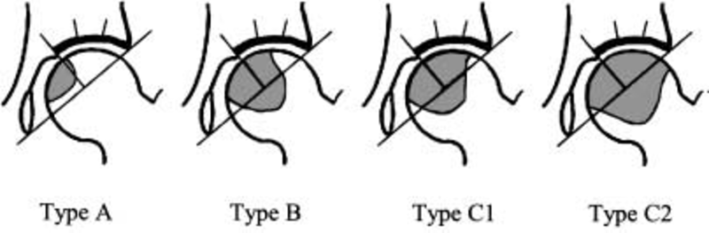
Fig. 1. 2001 revised classification of osteonecrosis. The classification scheme consists of four types (A, B, C1, and C2) and is based on the central coronal section of the femoral head on T1-weighted images or the anteroposterior x-ray view. Type A lesions occupy the medial one-third or less of the weightbearing portion. Type B lesions occupy the medial two-thirds or less of the weight-bearing portion. Type C1 and type C2 lesions both occupy more than the medial two-thirds of the weight-bearing portion, but, whereas type C2 lesions extend laterally to the acetabular edge, type C1 lesions do not. The weight-bearing portion is defined as the area lateral to the mid-vertical line of the line through the acetabular edge and the teardrop bottom.
图1. 2001 年修订的股骨头骨坏死分类示意图。分类方案由四种类型(A、B、C1和C2)组成,基于T1加权图像或前后位X线片上股骨头的冠状位中央部分。A型病变占据承重部分的内侧三分之一或更少。B型病变占据承重部分的内侧三分之二或更少。C1型和C2型病变均占据超过承重部分内侧三分之二的位置,但是,C2型病变横向延伸至髋臼边缘,而C1型病变则不然。承重部分定义为穿过髋臼边缘和泪滴底部的连线的中垂直线的外侧区域。
特发性股骨头坏死的诊断、分类和分期的2001年修订标准由日本厚生劳动省主持下的特定疾病调查委员会工作组于2001年6月提出,以制定特发性股骨头坏死的诊断和治疗标准。选择5个特异性较高的标准进行诊断:股骨头塌陷(包括新月征),X线图像上无关节间隙狭窄或髋臼异常;确定股骨头硬化,无关节间隙狭窄或髋臼异常;骨扫描中的“冷热”(图像异常);T1加权MRI上的低强度信号带(带状特征);以及组织学上的骨小梁和骨髓坏死。如果患者满足这五个标准中的两个并且没有骨肿瘤或发育不良,则诊断为特发性股骨头坏死。根据坏死病灶在T1加权图像或X线图像上的位置,将其分为四种类型。A型病变占据承重部分的内侧三分之一或更少。B型病变占据承重部分的内侧三分之二或更少。C1型病变占据承重部分内侧三分之二以上,但不横向延伸至髋臼边缘。C2型病变占据承重部分内侧三分之二以上,并横向延伸至髋臼边缘。分期基于X线图像上股骨头的前后位和侧位视图。第1阶段被定义为虽然在MRI、骨闪烁图或组织学上观察到具体结果,但X线图像上没有具体发现骨坏死的时期。第2阶段是观察到确定股骨头硬化而没有股骨头塌陷的时期。第3阶段是观察到股骨头塌陷(包括新月征)且关节间隙不狭窄的时期。第3阶段在股骨头或髋臼处可以观察到轻度骨赘形成。第3阶段分为两个亚阶段。在3A阶段,股骨头塌陷小于3毫米。在第3B阶段,股骨头塌陷为3毫米或更大。第四阶段是观察到骨关节炎变化的时期。
The 2001 revised criteria for diagnosis, classification, and staging of idiopathic osteonecrosis of the femoral head
Abstract
The 2001 revised criteria for the diagnosis, classification, and staging of idiopathic osteonecrosis of the femoral head were proposed in June 2001, by the working group of the Specific Disease Investigation Committee under the auspices of the Japanese Ministry of Health, Labor and Welfare, to establish criteria for diagnosis and management of idiopathic osteonecrosis of the femoral head. Five criteria that showed high specificity were selected for diagnosis: collapse of the femoral head (including crescent sign) without joint-space narrowing or acetabular abnormality on x-ray images; demarcating sclerosis in the femoral head without joint-space narrowing or acetabular abnormality; “cold in hot“ on bone scans; low-intensity band on T1-weighted MRI (bandlike pattern); and trabecular and marrow necrosis on histology. Idiopathic osteonecrosis of the femoral head is diagnosed if the patient fulfills two of these five criteria and does not have bone tumors or dysplasias. Necrotic lesions are classified into four types, based on their location on T1-weighted images or x-ray images. Type A lesions occupy the medial one-third or less of the weight-bearing portion. Type B lesions occupy the medial two-thirds or less of the weight-bearing portion. Type C1 lesions occupy more than the medial two-thirds of the weight-bearing portion but do not extend laterally to the acetabular edge. Type C2 lesions occupy more than the medial two-thirds of the weight-bearing portion and extend laterally to the acetabular edge. Staging is based on anteroposterior and lateral views of the femoral head on x-ray images. Stage 1 is defined as the period when there are no specific findings of osteonecrosis on x-ray images, although specific findings are observed on MRI, bone scintigram, or histology. Stage 2 is the period when demarcating sclerosis is observed without collapse of the femoral head. Stage 3 is the period when collapse of the femoral head, including crescent sign, is observed without joint-space narrowing. Mild osteophyte formation in the femoral head or acetabulum may be observed in stage 3. Stage 3 is divided into two substages. In stage 3A, collapse of the femoral head is less than 3 mm. In stage 3B, collapse of the femoral head is 3 mm or greater. Stage 4 is the period when osteoarthritic changes are observed.
文献出处:Nobuhiko Sugano, Takashi Atsumi, Kenji Ohzono, Toshikazu Kubo, Takao Hotokebuchi, Kunio Takaoka. The 2001 revised criteria for diagnosis, classification, and staging of idiopathic osteonecrosis of the femoral head. J Orthop Sci. 2002;7(5):601-5. doi: 10.1007/s007760200108.
本文是陶可版权所有,未经授权请勿转载。本文仅供健康科普使用,不能做为诊断、治疗的依据,请谨慎参阅
评论