学术前沿
发表者:马继光 人已读
中国医学科学院整形外科医院特需医疗部主任中国医学科学院整形外科医院整形科马继光
注射美容中心主任 马继光教授欢迎您
中国医学科学院整形外科医院特需医疗部
位于北京八大处的中国医学科学院整形外科医院是国内最负盛名的整形美容外科专科医院。
该院隶属于中国医学科学院和中国协和医科大学,是国内最早的整形外科学研究生培养基地,拥有40多名具有高级职称的教授和医师队伍,所培养的研究生和进修生遍布于全国各地。
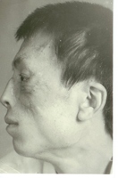
预扩张额部皮瓣鼻再造同时修复面部皮肤缺损
Simultaneous nasal reconstruction and facial defect repair using expanded forehead flap
MA Jiguang, YANG Xin
(Plastic Surgery Hospital,Peking Union Medical College, Chinese Academy of Medical Science,Beijing 100041,China.)
Abstract:ObjectiveTo improve the method for the repair of facial defect using expanded forehead flap.Methods10 patients were treated with the expanded forehead flap for total nose reconstruction and repair of facial defects. The expanded flap was divided two parts: the supratrochlear vessels for nasal reconstruction and the frontal branch of the superficial temporal vessels for facial defects.The periorbital or zygomatic area, the upper or lower lip. The defect of the forehead donor site was directly closed.ResultsNasal reconstruction and repair of facial defects were satisfactory in all patients.ConclusionNasal reconstruction and simultaneous repair of facial defects extand the scope of use of expanded forehead flap.
Key words:Plastic rhinoplasty; Surgery flaps
Expanded forehead flap is employed in classic nasal reconstruction to avoid secondary defect after the linear closure of the donors site . We , since Sept 1991, have extended the use of expanded forehead flap by achieving nasal reconstruction and facial defect repair simultaneously.
MATERIALS:
Ten patients (8 males and 2 females )presenting with nasal and facial defect were recruited to our hospital from Sept. 1991 to Oct.1999 . The age of the patients ranged from 14 to 35 . Among them, 3 were bitten by animals, 5 suffered from burn injury and 2 with congenital melanocytic nevus. Nasal defect were seen in all the patients , combined with defects in other facial areas (orbital , zygomatic , upper lip or lower lip etc.). The maximum size of facial defect was 6cm x 4cm ,the minimum was 2cm x 6cm . The first operation was performed under local anesthesia , implanting a 240-340 ml ,column-shaped expander in the forehead .In the second operation under general anesthesia, the expander was taken out, nasal reconstruction was done using the part of expanded forehead flap, an axial pattern flap that relies on perfusion from one literal supratrochlear artery. The other part of expanded flap was modified as an island pattern flap obtaining blood supply from the frontal branch of the superficial temporal artery on the other side and was employed to repair the defects in other facial areas. The maximum size of the flap used in nasal reconstruction and that used to repair other defects were 8.5cm x 10cm and 6cm x 4cm respectively ,the minimum was 6.5cm x 8cm and 2cm x 6cm . The flap’s donor site was closed in a linear fashion. The pedicle of the supratrochlear flap was incised 2 weeks later. In the follow-up period which lasted 1-2 years, all patients were satisfied with the final outcome .
METHOD:
1. Pre-operational design: We selected appropriate expanders after having carefully evaluated the size and shape of the nasal and facial defect. As it turned out , the volume of the expanders we selected were generally 240-340ml ,most of which were column shaped. We used Dopplers to detect the exact start point and tract of the arteries involved and left them accurately marked with mythlene blue. In the operation, we used the upper part of expanded flap to for transposition and the lower part for closure of the donor site.This is most important to ensure a smooth closure of the donor site and an hidden scar behind the hair line.
2. repairing of the defects: The incision was made 5-7cm long behind the hair line in the middle. The whole area of forehead skin was sufficiently undermined beneath the apeunorosis and musculi frontalis carefully avoiding damaging the axial arteries. The expander was inserted and the wound was closed by layers . Drainage was placed in order to prevent post-operation complications such as hematoma and wound cleft .
The secondary procedure was performed 1 week after expansion had been finished. We then took out the expander, one side of the expanded flap, supplied by supratrochlear artery, was used for nasal reconstruction .The other side that was modified into an island pattern flap supplied by the frontal branch of the superficial temporal artery was employed to repair the facial defect . In the whole process, great caution was made to protect the axial arteries and ensure a sufficient width of the pedicle. The donor site was closed using the lower part of the flap. The nasal frame was sculptured using a piece of cartilage collected from one of the patient’s ribs. The pedicle containing the supratrochlear artery was incised 2 weeks later .
DISSCUSION :
Nasal reconstruction using expanded forehead flaps is widely reported[1-5]. We advocate the method of achieving nasal reconstruction and repair of facial defect simultaneously in the cases of nasal defect combined with defects in other subunits of the face. The outcome turned out to be satisfying. On the count of the anatomical characteristics of the axial arteries, we choose larger expanders to obtain abundant extra-skin. One part of the flap is used for nasal reconstruction and the other for repair of defects in other subunits. This method substantially extend the use of expanded forehead flap and provide another alternative for facial defect repairing .
The pre-operational design of this procedure is quite complicated and a sophisticated operational skill is also demanded. Careful design and exquisite operating are both of critical importance to insure a satisfying outcome. Here , we would like to share some of our experiences : (1) Accurately evaluate the size and shape of the defects. We suggest larger expanders , mostly 340ml in our cases, meticulously exceeding the normal volume when expanding to meet the large demand of extra-skin. (2) Two axial pattern flaps are used in this procedure and we recommend a thorough study of anatomy of the arteries involved. We suggest the use of Doppler before and after expansion of the flap and mark the exact start point and tract of the axial arteries. We use the upper part of flap for transposition and lower part for wound closure in order to put the scar behind the hair line as far as possible. (3) Due to the large volume of expander employed, tiny clefts in the wound may be observed during the mid and late stage of expansion. According to our experience, the collagen capsule surrounding the expander may perform the role as an infection localizer and thus make the taking out not necessary. We suggest rinsing of the area using antibiotics and saline and carrying on expanding without detrimental effect to the accomplishment of the whole plan .
REFERENCES:
[1]张涤生,金一涛.皮肤软组织扩张术应用于烧伤晚期整复(附10例报告).中华整形烧伤外科杂志,1985,1:24-26.
[2]Adamson JE. Nasal reconstruction with the expanded forehead flap. Plast Reconstr Surg,1988,81:12-15.
[3]Bolton LL. Forehead expansion and total nasal reconstruction. Ann Plast Surg, 1988,21:210-213.
[4]鲁开化,汪良能,罗锦辉.扩张后的额部皮瓣行鼻再造术.中华整形烧伤外科杂志,1989,5:182.
[5]钟德才,鲁开化,艾玉峰.额部扩张皮瓣鼻再造.中华整形烧伤外科杂志, 1994,10:178-181.
本文是马继光版权所有,未经授权请勿转载。 本文仅供健康科普使用,不能做为诊断、治疗的依据,请谨慎参阅
发表于:2014-10-21